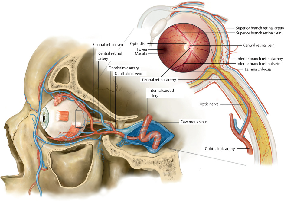
The retinal vascular occlusion refers to the network of blood vessels in the retina, a thin layer of tissue at the back of the eye. The retinal is responsible for capturing light and sending visual signals to the brain via the optic nerve. The retinal vascular system provides the retina with the necessary oxygen and nutrients to function correctly.
Structure of the Retinal Vascular Occlusion
Arteries: Arteries carry oxygenated blood from the heart to the retina. In the eye, they branch into smaller arterioles, which further divide into capillaries.
Veins: Veins carry deoxygenated blood away from the retina and back to the heart. Similarly, they branch into venules before returning to larger veins.
Types of Retinal Vascular Occulsion
Retinal vascular occlusion refers to the blockage of blood vessels that supply the retina, leading to a disruption in blood flow. This condition can cause severe visual impairment or even blindness if not promptly addressed. There are two main types of retinal vascular occlusion: central retinal vein occlusion (CRVO) and central retinal artery occlusion (CRAO). Here’s an explanation of each:
Illustrative image Central Retinal Vein Occlusion (CRVO)
Central Retinal Vein Occlusion (CRVO):
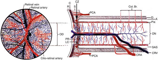
- Description: CRVO occurs when the central retinal vein, which drains blood from the retina, becomes blocked. This blockage leads to a buildup of blood and fluid in the retina, causing swelling (edema) and compromising the retinal function.
- Risk Factors: Hypertension, diabetes, glaucoma, atherosclerosis, blood clotting-disorders, and age-related factors are among the risk factors associated with CRVO.
- Symptoms: Sudden painless vision loss, blurry vision, distorted or missing areas in the visual field, and sometimes, floaters.
Illustrative image of Central Retinal Artery Occlusion (CRAO)
Central Retinal Artery Occlusion (CRAO):
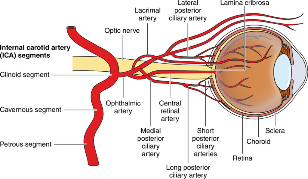
- Description: CRAO occurs when the central retinal artery, responsible for supplying oxygen and nutrients to the retina, becomes blocked. The lack of blood flow to the retina can lead to severe and rapid vision loss
- Risk Factors: Atherosclerosis, embolism (clot or debris), cardiac issues, giant cell arteritis, and other vascular diseases are commonly associated with CRAO.
- Symptoms: Sudden painless and severe vision loss, a “cherry-red spot” at the fovea (the central part of the retina), and a pale appearance of the optic nerve head
It’s important to note that both CRVO and CRAO are serious conditions that require immediate medical attention. Timely intervention can help minimize vision loss and, in some cases, may restore blood flow to the affected area. Treatment options may include medications, laser therapy, or surgery, depending on the underlying cause and severity of the occlusion. Regular eye exams and addressing underlying health conditions can also play a crucial role in preventing retinal vascular occlusions. If you suspect any issues with your vision, it is advisable to consult with an eye care professional for a thorough examination and appropriate management.
Symptoms of Retinal Vascular Occlusion
Symptoms related to the retinal vascular system can be indicative of various eye conditions, including:
1. Vision Changes: Blurred vision, loss of vision, or distortions in vision can occur when the retinal vessels are affected.
2. Floaters: The appearance of small, dark spots or specks floating in your field of vision.
3. Flashes of Light: Sudden flashes of light in your vision.
4. Bleeding or Hemorrhage: Blood in the eye can cause spots or streaks in your vision.
Effects of Retinal Vascular System
Disorders or conditions that affect the retinal vascular system can have significant consequences for vision and overall eye health. Some potential effects include:
1. Retinopathy: Damage to the retinal blood vessels, which can result in diabetic retinopathy, hypertensive retinopathy, or other types of retinal vascular diseases.
2. Macular Edema: Swelling of the macular, the central part of the retina responsible for detailed central vision.
3. Retinal Ischemia: Reduced blood flow to the retina can lead to retinal ischemia, which can cause vision loss or even blindness.
Causes of Retinal Vascular System
The causes of retinal vascular issues can vary depending on the specific condition:
1. Diabetes: Diabetic retinopathy is a common cause, where high blood sugar levels damage the retinal blood vessels.
2. Hypertension (High Blood Pressure): Hypertensive retinopathy can result from prolonged high blood pressure, leading to blood vessel damage in the retina.
3. Atherosclerosis: The buildup of plaque in the arteries can affect retinal blood flow.
4. Blood Clots: Blood clots in retinal blood vessels can block blood flow and cause retinal ischemia.
5. Other Conditions: Certain systemic diseases, like sickle cell anemia, and eye-specific conditions can also impact the retinal vascular system.
Treatments for Retinal Vascular System
Treatment for retinal vascular conditions depends on the specific diagnosis and underlying cause. Some common treatment options include:
1. Lifestyle Modifications: Managing underlying conditions like diabetes or hypertension through lifestyle changes and medication.
2. Laser Therapy: In some cases, laser therapy (laser photo-coagulation) may be used to seal leaking blood vessels or treat abnormal blood vessel growth.
3. Anti-VEGF Injections: Medications that block the action of vascular endothelial growth factor (VEGF) can be injected into the eye to treat conditions like macular edema.
4. Surgery: In advanced cases, surgical procedures may be required to repair or replace damaged blood vessels.
5. Regular Eye Exams: Early detection through routine eye exams is crucial for timely intervention.
It’s important to note that the prognosis and treatment options vary depending on the specific retinal vascular condition, its severity, and how promptly it is diagnosed and managed. Regular eye check-ups and managing underlying health conditions are essential for maintaining the health of the retinal vascular system and preserving vision.
RETINAL DISEASES
Retinal diseases encompass a diverse group of eye conditions that affect the retina, the light-sensitive tissue at the back of the eye responsible for capturing visual images and transmitting them to the brain via the optic nerve. Various factors, including genetics, age, systemic health, and environmental factors, can contribute to the development of retinal diseases. Here are some common retinal diseases:
Age-Related Macular Degeneration (AMD):
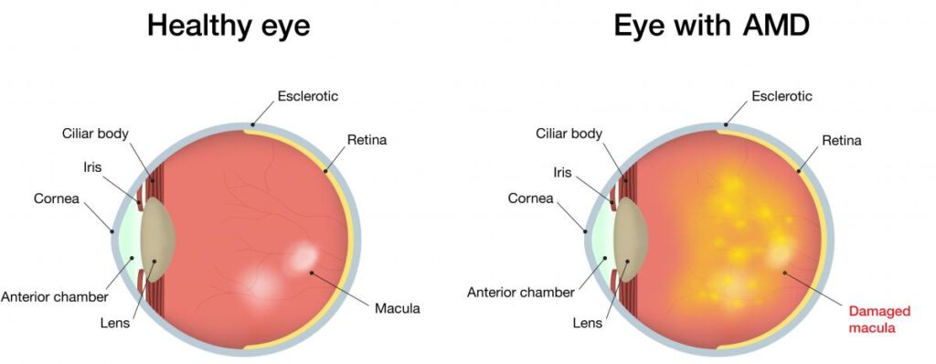
- Description: AMD is a progressive condition that primarily affects older adults. It involves the deterioration of the macula, the central part of the retina responsible for sharp, central vision.
- Symptoms: Blurred or distorted central vision, difficulty reading or recognizing faces, and the appearance of dark spots in the central vision.
Diabetic Retinopathy:
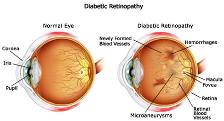
- Description: Diabetic retinopathy is a complication of diabetes that affects the blood vessels in the retina. High blood sugar levels can damage the blood vessels, leading to leakage, swelling, and abnormal growth.
- Stages: It can progress through stages, including non-proliferative diabetic retinopathy (mild, moderate, severe) and proliferative diabetic retinopathy, characterized by the growth of abnormal blood vessels.
Retinal Detachment:
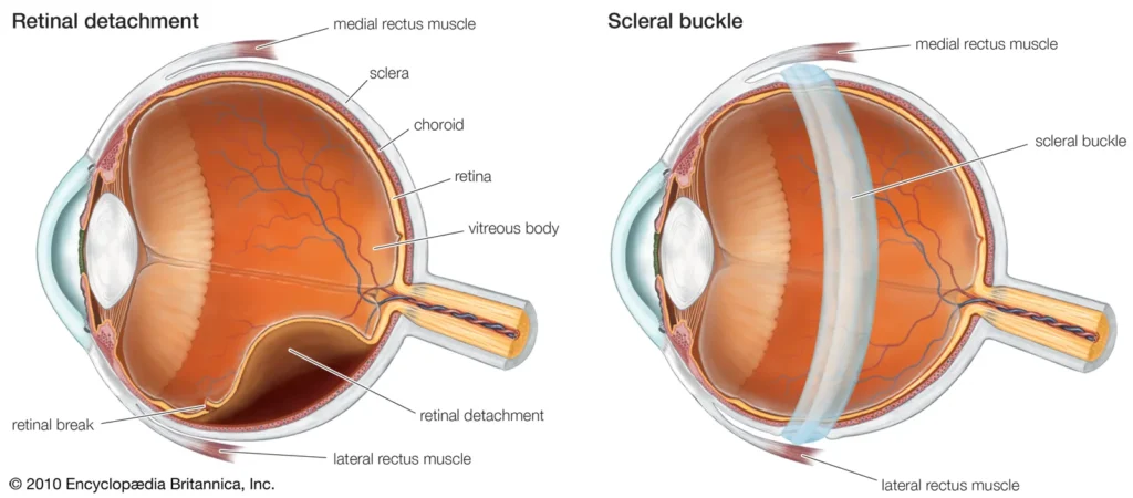
Description: Retinal detachment occurs when the retina pulls away from the underlying tissue. This can lead to a sudden onset of vision loss and is considered a medical emergency.
Causes: Trauma, aging, and conditions such as diabetic retinopathy can increase the risk of retinal detachment.
Retinitis Pigmentosa (RP):
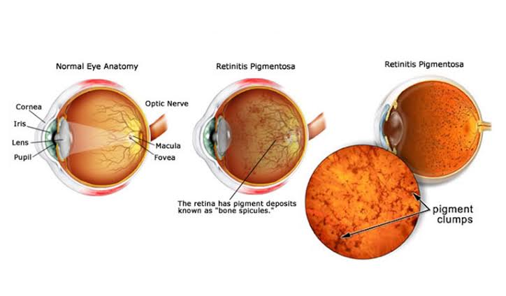
Description: RP is a group of inherited disorders that involve a breakdown and loss of cells in the retina, leading to progressive vision loss. It often starts with difficulty seeing in low-light conditions.
The most common early symptom of RP is loss of night vision — usually starting in childhood. Parents may notice that children with RP have trouble moving around in the dark or adjusting to dim light.
RP also causes loss of side (peripheral) vision — so you have trouble seeing things out of the corners of your eyes. Over time, your field of vision narrows until you only have some central vision (also called tunnel vision).
Some people with RP lose their vision more quickly than others. Eventually, most people with RP lose their side vision and their central vision.
Other symptoms of RP include:
- Sensitivity to bright light
- Loss of color vision
What causes RP?
Most of the time, RP is caused by changes in genes that control cells in the retina. These changed genes are passed down from parents to children.
RP is linked to many different genes and can be inherited in different ways. If you have RP, you can talk with your doctor or a specialist called a genetic counselor to learn more about your risk of passing RP to your children.
Sometimes RP happens as part of other genetic conditions, like Usher syndrome. Usher syndrome causes both vision and hearing loss.
RP can also be caused by some medicines, infections, or by an eye injury — but these causes aren’t as common.
Genetic Factors: RP is caused by various genetic mutations, and its severity can vary among individuals.
Retinal Vascular Diseases:
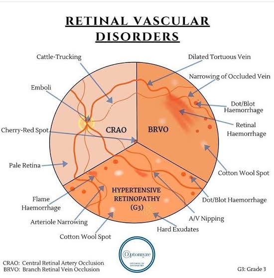
Description: These include conditions such as retinal vein occlusion (RVO) and retinal artery occlusion (RAO), which were briefly mentioned in the previous response.
Risk Factors: Hypertension, diabetes, and atherosclerosis are common risk factors for retinal vascular diseases.
Macular Holes and Epiretinal Membranes:

Description: Macular holes are small breaks in the macula, while epiretinal membranes are thin sheets of tissue that can form on the retina’s surface. Both can distort vision.
Symptoms: Central vision distortion, straight lines appearing wavy, and decreased central vision clarity.
Treatment for retinal diseases varies depending on the specific condition but may include medications, laser therapy, surgery (such as vitrectomy), and lifestyle modifications. Regular eye examinations, especially for individuals with risk factors, are crucial for early detection and management of retinal diseases. If you experience any changes in your vision, it’s essential to seek prompt evaluation by an eye care professional.
CONCLUSION
“In navigating the complexities of Retinal Vascular Occlusion, it is imperative to recognize the unique challenges faced by individuals in the African context. Access to specialized healthcare resources, awareness, and culturally sensitive support are pivotal in addressing the impact of this condition. Together, as a united healthcare community, let us strive to bridge gaps, promote education, and empower individuals affected by Retinal Vascular Occlusion in Africa. By fostering collaboration, understanding, and tailored interventions, we can enhance the quality of care and contribute to a brighter future for those grappling with this condition on the African continent.”


