Introduction
Renal vein stenosis often emerges as a sequelae of various pathological processes, including atherosclerosis, fibromuscular dysplasia, and thrombosis, which collectively contribute to the narrowing of the renal veins. The clinical spectrum of renal vein stenosis ranges from asymptomatic cases to severe hypertension and renal insufficiency. Accurate diagnosis proves challenging due to the subtle nature of early symptoms and the diverse array of potential underlying causes.
This article explores the multifaceted landscape of renal vein stenosis, shedding light on the nuanced interplay between vascular dynamics and renal function. By examining the latest advancements in imaging techniques, such as Doppler ultrasound and magnetic resonance angiography, clinicians can refine their diagnostic acumen and tailor interventions to individual patient needs. Moreover, advancements in endovascular procedures and surgical interventions have reshaped the therapeutic landscape, offering novel avenues for managing this intricate vascular condition.
As we delve into the intricate web of factors influencing renal vein stenosis, this exploration aims to equip healthcare professionals with a comprehensive understanding of the disorder, fostering improved clinical decision-making and patient outcomes in the challenging realm of renal vascular pathology.
RENAL VEIN STENOSIS (RVS)/RENAL ARTERY STENOSIS (RAS)
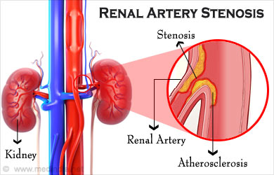
Renal Vein Stenosis also known as renal artery stenosis, is a medical condition characterized by the narrowing or constriction of one or both renal veins. These veins carry deoxygenated blood from the kidneys back to the heart. When they become narrowed, it can disrupt normal blood flow and have significant consequences for kidney function and overall health.
Causes of Renal Vein Stenosis
1. Atherosclerosis: The most common cause of RVS is atherosclerosis, a condition in which fatty deposits, cholesterol, and other substances build up on the walls of blood vessels, including the renal arteries and veins. This buildup can narrow the renal vein.
2. Fibro-muscular Dysplasia (FMD): FMD is a rare condition in which the walls of the renal artery (and sometimes the renal vein) develop abnormally. This can result in stenosis of the renal vein.
3. Blood Clots: Blood clots can form in the renal veins, either directly or as a result of other conditions like deep vein thrombosis (DVT). These clots can obstruct blood flow.
Effects of Renal Vein Stenosis
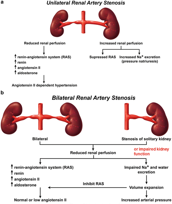
1. Hypertension (High Blood Pressure): RVS can lead to high blood pressure, a condition known as renovascular hypertension. The kidneys play a crucial role in regulating blood pressure, so any disruption in renal blood flow can lead to hypertension. Reno-vascular hypertension is high blood pressure due to narrowing of the arteries that carry blood to the kidneys. This condition is also called renal artery stenosis.
Causes
Renal artery stenosis is a narrowing or blockage of the arteries that supply blood to the kidneys.
The most common cause of renal artery stenosis is a blockage in the arteries. This problem most often occurs when a sticky, fatty substance called plaque builds up on the inner lining of the arteries, causing a condition known as atherosclerosis.
When the arteries that carry blood to your kidneys become narrow, less blood flows to the kidneys. The kidneys mistakenly respond as if your blood pressure is low. As a result, they release hormones that tell the body to hold on to more salt and water. This causes your blood pressure to rise.
Risk factors for atherosclerosis:
- High blood pressure
- Smoking
- Diabetes
- High cholesterol
- Heavy alcohol use
- Cocaine abuse
- Increasing age
Fibromuscular dysplasia is another cause of renal artery stenosis. It is typically seen in women under age 50. It tends to run in families. The condition is caused by abnormal growth of cells in the walls of the arteries leading to the kidneys. This also leads to narrowing or blockage of these arteries.
Symptoms
People with renovascular hypertension may have a history of very high blood pressure that is hard to bring down with medicines.
Symptoms of renovascular hypertension include:
- High blood pressure at a young age
- High blood pressure that suddenly gets worse or is hard to control
- Kidneys that are not working well (this can start suddenly)
- Narrowing of other arteries in the body, such as to the legs, the brain, the eyes and elsewhere
- Sudden buildup of fluid in the air sacs of the lungs (pulmonary edema)
If you have a dangerous form of high blood pressure called malignant hypertension, symptoms can include:
- Bad headache
- Nausea or vomiting
- Confusion
- Changes in vision
- Nosebleeds
Exams and Tests
The health care provider may hear a “whooshing” noise, called a bruit, when placing a stethoscope over your belly area.
The following blood tests may be done:
- Cholesterol levels
- Renin and aldosterone levels
- BUN (blood urea nitrogen)
- Creatinine
- Potassium
- Creatinine clearance
Imaging tests may be done to see if the kidney arteries have narrowed. They include:
- Angiotensin converting enzyme (ACE) inhibition renography
- Doppler ultrasound of the renal arteries
- Magnetic resonance angiography (MRA)
- Renal artery angiography
Treatment
High blood pressure caused by narrowing of the arteries that lead to the kidneys is often hard to control.
One or more medicines are needed to help control blood pressure. There are many types available.
- Everyone responds to medicine differently. Your blood pressure should be checked often. The amount and type of medicine you take may need to be changed from time to time.
- Ask your provider what blood pressure reading is right for you.
- Take all medicines the way your provider prescribed them.
Have your cholesterol levels checked, and treated if it is needed. Your provider will help determine the right cholesterol levels for you based on your heart disease risk and other health conditions.
Lifestyle changes are important:
- Eat a heart-healthy diet.
- Exercise regularly, at least 30 minutes a day (check with your doctor before starting).
- If you smoke, quit. Find a program that will help you stop.
- Limit how much alcohol you drink: 1 drink a day for women, 2 a day for men.
- Limit the amount of sodium (salt) you eat. Aim for less than 1,500 mg per day. Check with your doctor about how much potassium you should be eating.
- Reduce stress. Try to avoid things that cause stress for you. You can also try meditation or yoga.
- Stay at a healthy body weight. Find a weight-loss program to help you if you need it.
Further treatment depends on what causes the narrowing of the kidney arteries. Your provider may recommend a procedure called angioplasty with stenting.
These procedures may be an option if you have:
- Severe narrowing of the renal artery
- Blood pressure that cannot be controlled with medicines
- Kidneys that are not working well and are becoming worse
However, the decision about which people should have these procedures is complex, and depends on many of the factors listed above.
Possible Complications
If your blood pressure is not well controlled, you are at risk for the following complications:
- Aortic aneurysm
- Heart attack
- Heart failure
- Chronic kidney disease
- Stroke
- Vision problems
- Poor blood supply to the legs
When to Contact a Medical Professional
Contact your provider if you think you have high blood pressure.
Contact your provider if you have renovascular hypertension and symptoms get worse or do not improve with treatment. Also call if new symptoms develop.
Prevention
Preventing atherosclerosis may prevent renal artery stenosis. Taking the following steps can help:
- Lose weight if you are overweight.
- Ask your provider about your smoking and alcohol use.
- Control your blood sugar if you have diabetes.
- Make sure your provider is monitoring your blood cholesterol levels.
- Eat a heart-healthy diet.
- Get regular exercise.
2. Kidney Dysfunction: Reduced blood flow in the renal veins can impair kidney function. This can result in decreased filtration of waste products and excess fluids, potentially leading to conditions like chronic kidney disease (CKD) or kidney failure.
Kidney Disease and Renal Failure
Kidney Disease resulting in Renal Failure is the malfunction of the kidneys causing the kidneys to lose their filtering ability. As a result of kidney disease, kidney failure or kidney injury, waste accumulates in the body and the body’s chemical balance gets disrupted. Symptoms of Kidney Disease and Renal Failure include fluid retention, fatigue, shortness of breath and decreased urine output.
Kidney Disease and Renal Failure Dialysis
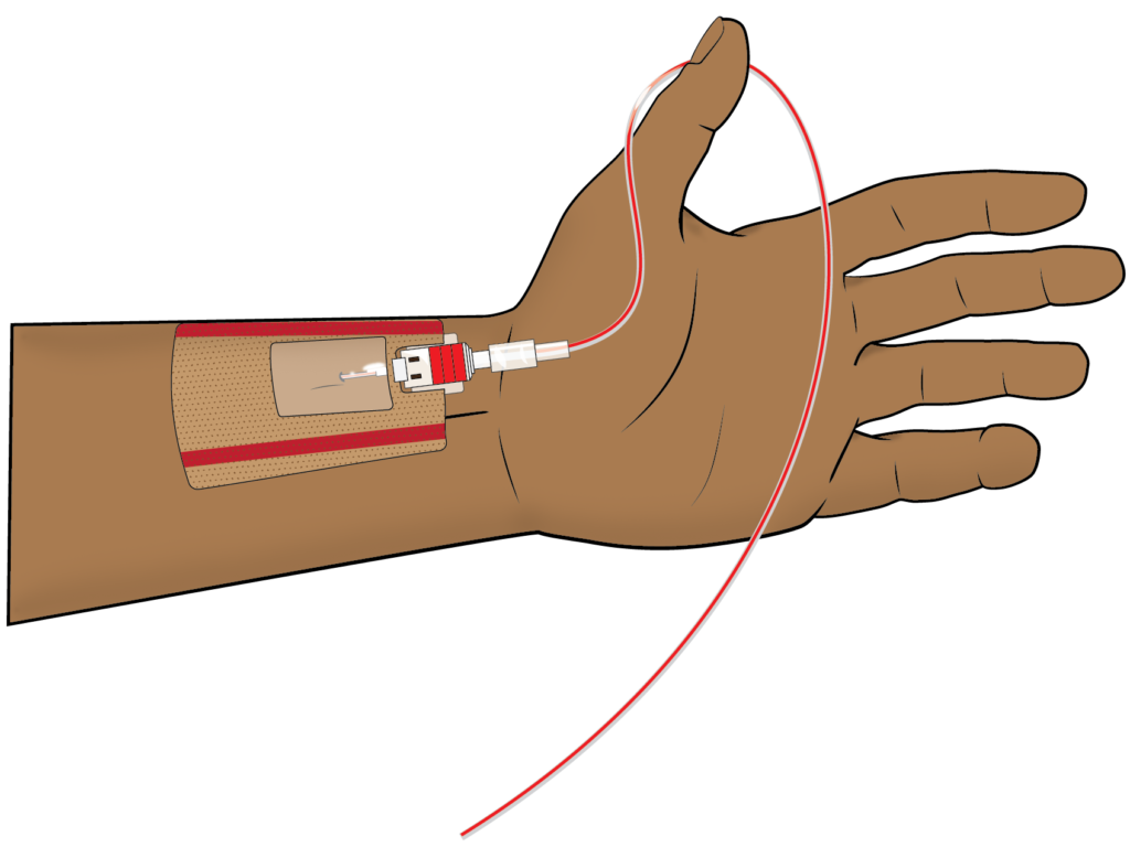
AV Fistulas are often the preferred route for hemodialysis treatment plans. These access routes are limited so vein preservation is important. Vascular Wellness makes vein preservation part of every clinician’s assessment.
Symptoms of Renal Artery Stenosis
The symptoms of RVS can vary widely, and some individuals may not experience noticeable symptoms. Common symptoms include:
1.Hypertension: High blood pressure is a frequent symptom, often resistant to standard anti-hypertensive medications.
2.Flank Pain: Some individuals may experience abdominal or flank pain, especially on the side of the affected kidney.
3.Decreased Kidney Function: This can lead to symptoms such as reduced urine output, swelling (edema), and elevated levels of waste products in the blood.
Diagnosis of Renal Artery Stenosis
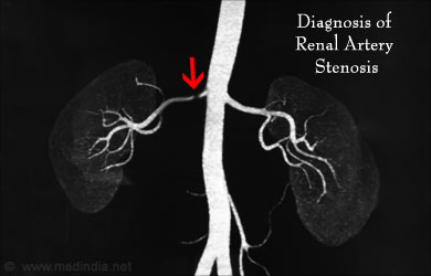
Auscultation usually provides initial clues about the presence of RAS. When blood flows through a narrow artery it usually makes a “whooshing” sound called a bruit. Doctors usually follow-up with other diagnostic imaging tests like:
1. Duplex ultrasound – which combines traditional ultrasound with Doppler ultrasonography. This is a painless, non-invasive technique that does not require anesthesia.
2. Catheter angiogram – is a type of x-ray where a thin, flexible tube called catheter is threaded through the large arteries from the groin to the renal artery. A contrast dye is injected through the catheter to view the renal artery clearly. This is a technique requiring a sedative and usually performed as an outpatient.
3. Computerized tomographic angiography (CTA) scan – which is a combination of x-rays and computer technology to create high-resolution x-rays. A contrast dye is injected in the patient’s vein to enable a clearer view of the arteries. This is again a non-invasive, outpatient procedure.
4. Magnetic resonance angiogram (MRA) – this technique uses radio waves and magnets to create detailed images of internal organs. Again a contrast dye may be used. This is also a non-invasive, outpatient procedure.
Treatment of Renal Vein/Artery Stenosis
Treatment for RVS/RAS aims to alleviate symptoms, improve kidney function, and manage associated conditions. The choice of treatment depends on the underlying cause and the severity of the stenosis. Treatment options include:
LIFESTYLE CHANGES – including quitting smoking, reducing alcohol, low-fat diet and adequate exercise. Patients with RVS can benefit from making certain lifestyle changes to help manage their condition and maintain overall health;
a. Diet: A diet low in sodium (salt) and high in fruits, vegetables, and whole grains can help manage blood pressure and reduce strain on the kidneys.
b. Exercise: Regular physical activity can aid in controlling blood pressure and improving overall cardiovascular health.
c. Smoking Cessation: If the patient smokes, quitting smoking is highly recommended, as smoking can worsen the effects of RVS on blood vessels.
d. Medication Adherence: It’s essential to take prescribed medications consistently and as directed by your healthcare provider to control blood pressure and protect kidney function.
MEDICATIONS – is aimed at keeping RVH in check. Patients may be prescribed either an Angiotensin-converting enzyme (ACE) inhibitors or angiotensin receptor blockers (ARBs). Diuretics are also helpful in aiding the kidneys remove fluid from the blood. Beta-blockers and calcium-channel blockers and other anti-hypertensive drugs may be required. In some cases, a cholesterol lowering medication and blood thinners may be required.
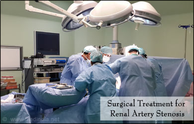
SURGERY – is usually recommended for patients with RAS who do not improve with medications. Surgery options include angioplasty and stenting where a catheter is inserted into the renal artery and a mesh tube (stent) is placed to keep the plaque flattened and the artery open.
Another surgical method is known as bypass surgery where a bypass is created with a vein or synthetic tube to connect the kidney to the aorta. This creates an alternate route for blood flow around the blocked artery into the kidney. An endarterectomy is a method where the plaque is cleaned from the artery thereby clearing it for smooth blood flow.
Treatment options and plans are usually decided on a case-to-case basis by the treating physician, nephrologist and surgeon.
Risk Factors for Renal Artery Stenosis
Understanding the risks and taking precautions is the best way to prevent complications from RAS. Some of the risk factors include:
1. Age – over 50 years
2. Diabetes
3. High blood pressure
4. Cholesterol
5. Smoking
6. Alcohol
7. Family history of coronary heart disease or peripheral arterial disease
8. Family history of RAS
Outlook:
The prognosis for individuals with RVS depends on various factors, including the cause, the extent of stenosis, and how promptly it’s diagnosed and treated. With appropriate medical care, lifestyle modifications, and regular monitoring, many people can effectively manage RVS and its complications. However, untreated or poorly managed RVS can lead to severe kidney damage and uncontrolled hypertension, highlighting the importance of early diagnosis and treatment.
However, it’s important to note that untreated or poorly managed RVS can lead to severe complications, including kidney damage and uncontrolled hypertension. This makes early diagnosis and treatment critical for a better long-term outlook.
If you or someone you know is experiencing symptoms suggestive of renal vein stenosis or has risk factors for the condition, it’s essential to seek medical evaluation and guidance. A healthcare provider can perform the necessary tests, such as imaging studies and blood pressure monitoring, to determine the diagnosis and develop an appropriate treatment plan.
FOLLOW-UP AND MONITORING:
After treatment for RVS, regular follow-up appointments with a healthcare provider are crucial to monitor kidney function and blood pressure. These visits help ensure that the treatment is effective and that any complications are promptly addressed.


